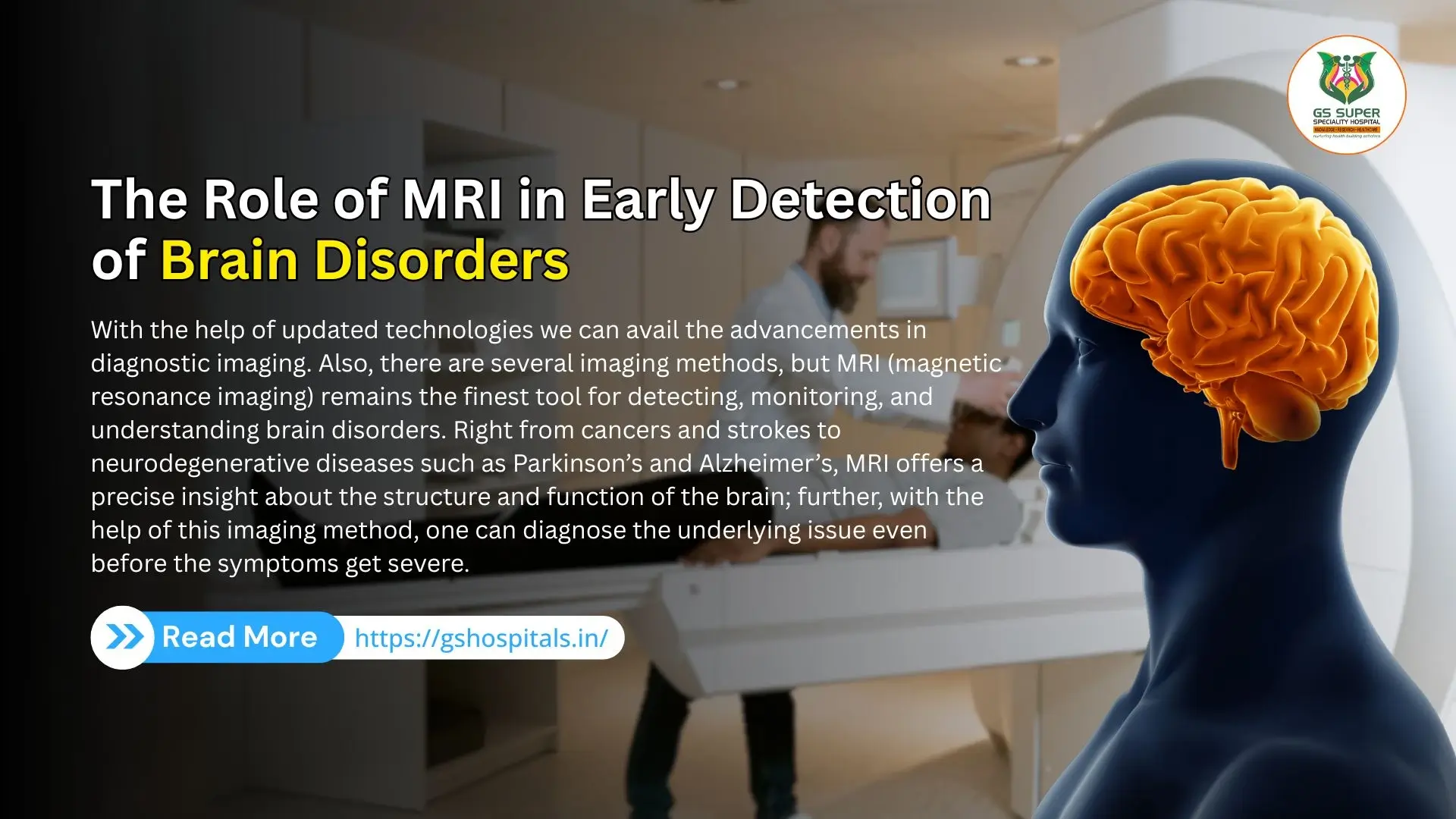
With the help of updated technologies we can avail the advancements in diagnostic imaging. Also, there are several imaging methods, but MRI (magnetic resonance imaging) remains the finest tool for detecting, monitoring, and understanding brain disorders. Right from cancers and strokes to neurodegenerative diseases such as Parkinson’s and Alzheimer’s, MRI offers a precise insight about the structure and function of the brain; further, with the help of this imaging method, one can diagnose the underlying issue even before the symptoms get severe. Let’s dive deeper into this blog to explore the significant role of MRI in diagnosing brain disorders on time and why it is considered a cornerstone in modern neurology.
As we all know MRI is a non-invasive imaging method, which uses highly efficient magnets and radio active waves to provide precise images of the particular vital organs and tissues in the body. MRI is one of the safer options to use because it won't employ any ionizing radiations such as CT scans and X-rays.
During the MRI scan, healthcare providers will employ the strong magnetic field to align the hydrogen atoms in the patient’s body. Further, this alignment will get disturbed by radiofrequency pulses, and the machine detects the energy released when atoms get back to their original position. This energy release is converted into high-resolution images. In the brain imaging, MRI offers:
Brain disorders such as degenerative, developmental, vascular in nature, or traumatic can eventually result in complex consequences if overlooked. Early detection will keep your health on better position, by:
MRI plays a critical role in diagnosing health issues in the brain even before they cause noticeable signs. Further, let’s explore how MRI is actually used for different brain disorders.
MRI detects brain activity primarily through a specialized technique known as functional Magnetic Resonance Imaging (fMRI). Unlike standard MRI scans for the brain, which provides detailed images of brain structures, fMRI measures changes in blood flow to different parts of the brain, which reflect neural activity.
When a specific area of the brain is more active, such as during thinking, movement, or seeing, neurons in that region require more oxygen. fMRI detects this change by measuring the Blood Oxygen Level Dependent (BOLD) signal, which tracks variations in oxygen-rich versus oxygen-poor blood. Oxygenated and deoxygenated hemoglobin have different magnetic properties, and fMRI captures these subtle differences.
This technique allows researchers and clinicians to generate real-time maps of brain activity while a person performs specific tasks, thinks, or even rests. fMRI is widely used in cognitive neuroscience research and clinical settings to identify regions responsible for language, memory, motor control, and more, especially useful in planning brain surgeries.
An MRI scan for the brain (Magnetic Resonance Imaging) is a non-invasive diagnostic tool used to produce detailed images of the brain’s structures. Its main purpose is to help doctors detect, diagnose, and monitor a wide range of neurological conditions. Unlike CT scans or X-rays, MRI uses powerful magnets and radio waves without radiation to create high-resolution images of soft tissues, making it especially valuable for examining the brain.
The Best Neurologist in Hapur, like those at GS hospital, will recommend an MRI scan for the brain for several reasons: it helps in the early detection of neurological disorders, evaluating brain injuries, diagnosing tumors, and monitoring chronic conditions..
In some cases, contrast dye (gadolinium) may be used during MRI to enhance image clarity, especially when looking for tumors, inflammation, or blood vessel issues.
Overall, the purpose of a brain MRI is to give a clear, accurate view of what’s happening inside the brain, allowing for early detection, targeted treatment, and better management of neurological health.
MRI plays a huge role in diagnosing structural changes of the brain on time, notably that linked with Alzheimer’s disease. It detects:
Volumetric MRI greatly helps in measuring brain atrophy and monitoring the progression of disease. Timely intervention in Alzheimer’s will assist in slowing down the cognitive decline with the help of medications, therapy, and lifestyle modifications.
Parkinson’s disease will be often detected though clinical symptoms, advanced MRI methods such as diffusion tensor imaging (DTI) and susceptibility-weighted imaging (SWI) can detect:
This supported the early detection of brain disorders and differentiation from similar movement disorders.
MRI takes a front seat when diagnosing brain cancers. It assists in:
Contrast-enhanced MRI assist will be precise visualization of tumors and tissues surrounded by it. fMRI (functional MRI) also assists in mapping brain areas near the tumor that are responsible for a few functions such as speech or movement.
MRI will easily diagnose the symptoms of stroke within a several hours of onset. DWI (diffusion-weighted imaging) is highly sensitive to timely ischemic changes, finding the disturbed brain tissue before it causes irreversible damage. This is crucial because stroke treatments such as thrombolysis are time-sensitive.
MRI will potentially find the difference between the various brain bleeds and pinpoint the source. SWI (susceptibility-weighted imaging) is ideal for detecting microbleeds and vascular malformations that surges the risk of stroke.
MRI remains the reliable source to diagnose and monitor Multiple Sclerosis, which is one of the chronic autoimmune diseases that disturbs the brain and spinal cord. MRI detects:
Regular MRIs assist in guiding therapy decisions and observe responses during the treatment.
While CT is used for emergency TBI assessment, MRI is far more sensitive to microstructural injuries, particularly in mild or moderate TBI where CT may be normal.
MRI reveals:
MRI helps in predicting long-term cognitive or emotional outcomes after a brain injury.
For individuals with epilepsy, especially drug-resistant epilepsy, MRI plays a key role in:
High-resolution MRI can detect subtle lesions that may be missed on routine imaging.
MRI can assist in evaluating children with developmental delays, intellectual disability, or Autism Spectrum Disorder (ASD) by identifying:
This aids in understanding the cause, guiding therapies, and offering prognosis.
Though psychiatric disorders are diagnosed clinically, research-level MRIs have shown promise in revealing structural and functional changes in:
In the future, MRI biomarkers may aid in personalized treatment for mental health disorders.
Despite its many advantages, MRI has a few limitations:
However, as technology advances, many of these limitations are being addressed through faster sequences, open MRI scanners, and better patient preparation.
Artificial intelligence (AI) and machine learning are enhancing MRI capabilities by:
In the near future, MRI may also be used in preventive neurology, screening high-risk individuals (family history, genetic predisposition) to catch diseases before symptoms appear.
One of its biggest strengths is its ability to produce high-resolution images of the brain's soft tissues, allowing the best neurology doctor in Hapur like those at GS hospital, to detect abnormalities with great precision. GS super specialty Hospital is known for its advanced diagnostic facilities and expert neurology care.
A major benefit of MRI is that it’s a non-invasive and radiation-free procedure. Unlike CT scans or X-rays, MRI does not use ionizing radiation, making it safer, especially for repeated imaging or for vulnerable groups like children and pregnant women (with caution).
MRI is highly effective in detecting a wide range of brain issues, including tumors, stroke, multiple sclerosis, infections, aneurysms, and traumatic injuries. Advanced MRI techniques such as fMRI, diffusion tensor imaging, and MR spectroscopy provide detailed insights into brain function, blood flow, and even chemical composition.
Another significant advantage is the use of contrast agents like gadolinium, which further enhance image clarity and help distinguish between different types of tissues or abnormalities.
In summary, the pros of an MRI brain scan include superior image quality, safety, versatility, and functional imaging capabilities, making it an essential tool in modern neurology for early detection, diagnosis, and treatment planning.
MRI is generally considered a very safe and non-invasive imaging technique, and it does not cause any harm or damage to the brain. It uses strong magnetic fields and radiofrequency waves to create detailed images of brain structures without exposing the body to ionizing radiation, which is a key advantage over X-rays or CT scans.
There is no scientific evidence to suggest that MRI itself causes brain tissue damage, memory problems, or neurological issues. The magnetic fields and radio waves used in MRI are not strong enough to alter brain cells or brain function.
However, certain precautions are necessary. People with metal implants, pacemakers, aneurysm clips, or cochlear implants may be at risk because the magnetic field can interfere with these devices. Also, the confined space and loud noises during the scan can cause discomfort, anxiety, or claustrophobia in some individuals.
In rare cases, if a contrast dye (gadolinium) is used, it might cause allergic reactions or mild side effects like headache or nausea. Gadolinium is usually safe, but it should be avoided in patients with severe kidney issues.
Overall, MRI does not cause any direct issues in the brain and is widely used due to its safety and accuracy.
1. What is the purpose of a brain MRI?
To detect, diagnose, and monitor brain disorders with high-detail imaging.
2. Can MRI detect early brain changes?
Yes, MRI can reveal abnormalities before symptoms appear.
3. Is MRI safe for repeated use?
Yes, it is radiation-free and generally safe for most individuals.
4. Which conditions can MRI detect early?
Alzheimer’s, tumors, stroke, multiple sclerosis, and more.