
Brain Hemorrhage: Causes, Symptoms, and Emergency Treatment
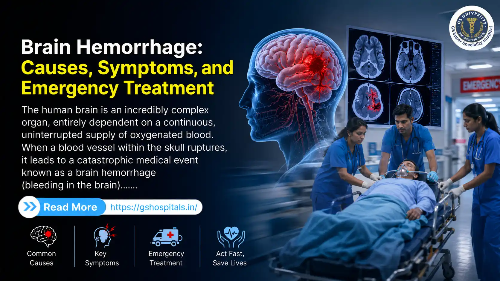
The human brain is an incredibly complex organ, entirely dependent on a continuous, uninterrupted supply of oxygenated blood. When a blood vessel within the skull ruptures, it leads to a catastrophic medical event known as a brain hemorrhage (bleeding in the brain). This localized bleeding kills brain cells in two ways: by cutting off downstream oxygen delivery and by creating localized tissue pressure (a hematoma) inside the rigid, unyielding confines of the skull structure.
Because brain cells begin dying within minutes of oxygen deprivation, recognizing brain hemorrhage symptoms early and choosing a leading stroke treatment hospital can mean the difference between full recovery and permanent neurological damage. Seeking an expert brain hemorrhage specialist at a highly advanced neurosurgery hospital is paramount to survival and recovery.
Primary Causes of a Brain Hemorrhage
A ruptured blood vessel in the brain rarely occurs without an underlying vascular weakness, systemic disease, or physical impact. Understanding these root causes can aid in preventative health measures and risk reduction.
Chronic Hypertension (High Blood Pressure)
Hypertension is the single most common, modifiable risk factor for a non-traumatic brain hemorrhage. Over decades, untreated or poorly managed high blood pressure exerts constant mechanical stress on the walls of small, deep arteries within the brain.
Head Trauma
Physical trauma to the head is the primary cause of intracranial bleeding in young adults and children, though it is also heavily prevalent among the elderly due to falls.
Cerebral Aneurysms
An aneurysm is a structural defect where a localized portion of an artery wall balloons outward due to weakness in the vessel's muscular layer. These look like small berries hanging from a vascular fork.
Cerebral Amyloid Angiopathy (CAA)
Often observed in older adults (typically over the age of 65), cerebral amyloid angiopathy is a condition where abnormal protein deposits, called amyloid proteins, build up inside the walls of the brain’s small-to-medium-sized arteries.
Arteriovenous Malformations (AVMs)
An AVM is a congenital abnormality consisting of an abnormal, tangled web of blood vessels where arteries connect directly to veins without an intervening network of capillaries. Capillaries normally slow down blood flow and lower its pressure before it reaches the fragile veins. Without them, high-pressure arterial blood flows directly into thin-walled veins, causing them to stretch, dilate, and eventually rupture. AVMs are usually silent until they bleed, often occurring in teenagers and young adults.
Bleeding Disorders and Anticoagulants
Conditions that impair the body's natural ability to form blood clots—such as hemophilia, advanced liver disease, or severe thrombocytopenia (low platelet count)—substantially increase the risk of a brain bleed. Furthermore, the widespread use of prescription blood thinners (anticoagulants and antiplatelet drugs) for heart conditions or stroke prevention can turn a minor, otherwise negligible vascular leak into a massive, life-threatening brain hemorrhage.
Recognizing Brain Hemorrhage Symptoms
Symptoms can appear abruptly within seconds, or they can evolve gradually over several hours as a slow venous leak builds volume.
Recognizing these symptoms immediately is vital. If you or someone near you experiences these symptoms, prioritizing transit to a specialized neurosurgery hospital can save a life.
QUICK ACTION CHECKLIST (FAST+):
F - Face Drooping: Is one side of the face numb or sagging?
A - Arm Weakness: Ask them to raise both arms. Does one arm drift downward?
S - Speech Difficulty: Is their speech slurred, muddled, or hard to understand?
T - Time to Call Emergency: Note the onset time and seek immediate medical help.
+ - Sudden Severe Headache: Watch for an instant, blinding "thunderclap" headache.
The Sudden "Thunderclap" Headache
The most definitive symptom of a subarachnoid brain hemorrhage is a sudden, blinding headache of maximum intensity. Patients often describe this experience as a "thunderclap" because it hits out of nowhere like a crack of thunder. It is distinct from a typical migraine or tension headache due to its instantaneous onset and sheer severity.
Focal Neurological Deficits
Because different areas of the brain control different bodily functions, a hemorrhage in a specific region will cause localized signs:
- Hemiparesis or Hemiplegia: Sudden weakness, numbness, or total paralysis affecting only one side of the body (e.g., the left arm and left leg).
- Facial Asymmetry: One side of the mouth droops when the person tries to smile, or they lose control over their eyelid.
- Loss of Sensation: A sudden pins-and-needles sensation or complete numbness in a limb.
Speech and Visual Disturbances
The brain's language and visual processing centers are highly sensitive to pressure and oxygen loss:
- Aphasia: Complete inability to speak, slurred speech, or difficulty understanding spoken words. The person may speak in fragmented, nonsensical sentences.
- Visual Losses: Sudden blurring of vision, double vision (diplopia), or a complete loss of vision in one or both eyes. An asymmetric dilation of the pupils (where one pupil is vastly larger than the other) is a sign of high intracranial pressure compressing the cranial nerves.
Cognitive and Balance Disruptions
As pressure mounts globally within the skull, general brain function deteriorates rapidly:
- Ataxia: A sudden loss of balance, lack of coordination, and an inability to walk straight or remain upright.
- Altered Mental Status: Profound confusion, disorientation regarding time and place, extreme lethargy, or difficulty waking up. If left unmanaged, this quickly progresses to stupor and full coma.
- Seizures and Vomiting: Sudden, unprovoked seizures in someone without an epileptic history are common during a brain bleed. Sudden, projectile vomiting without any preceding nausea is another sign of rapidly escalating intracranial pressure.
Time is Brain: The Emergency Treatment Protocol
In the realm of neurology, the phrase "Time is Brain" is an absolute reality. When a blood vessel ruptures, approximately two million brain cells die every single minute that the bleeding continues uncontrolled and the pressure remains unmitigated. Managing a brain hemorrhage requires a highly coordinated, multi-phased medical response that begins the moment emergency services are contacted.
Phase 1: Immediate Pre-Hospital Stabilization
While waiting for medical professionals to arrive, follow these guidelines:
- Positioning: Keep the patient lying flat with their head and shoulders slightly elevated at approximately a 30-degree angle. This position utilizes gravity to help reduce venous pressure inside the head.
- Airway Monitoring: If the patient is unconscious or vomiting, gently roll them onto their side into the recovery position to prevent them from choking or aspirating fluids into their lungs.
- Zero Medications: Do not give the patient anything to eat or drink. Crucially, do not administer aspirin.
Phase 2: Hyper-Acute Diagnostic Imaging
The moment the patient arrives at the emergency department of a stroke treatment hospital, the medical team initiates emergency triage. The primary objective is to differentiate between an ischemic stroke and a hemorrhagic stroke, as their treatments are completely opposite.
The definitive diagnostic tool is a non-contrast Computed Tomography (CT) scan of the brain. A CT scan can detect fresh blood within minutes, appearing as a bright white mass against the darker gray tones of normal brain tissue. In some instances, a Magnetic Resonance Imaging (MRI) scan or a CT Angiogram (CTA) may be performed to locate the exact vessel that ruptured, mapping out any underlying aneurysms or AVMs.
Phase 3: Acute Medical Management
If the hemorrhage is small or located in a deeply delicate area where surgery would cause too much structural damage, the brain hemorrhage specialist will opt for aggressive medical stabilization:
- Blood Pressure Control: Intravenous medications (such as labetalol or nicardipine) are titrated to lower the patient's blood pressure into a safe range, slowing down the force of blood pumping out of the ruptured vessel.
- Reversal of Anticoagulation: If the patient was taking prescribed blood thinners, specific reversal agents (such as Vitamin K, protamine, or advanced clotting factor concentrates) are infused immediately to restore the blood's natural clotting ability.
- Intracranial Pressure (ICP) Control: Specialized osmotic diuretics, such as mannitol or hypertonic saline solutions, are administered intravenously to draw excess water out of healthy brain tissue and into the bloodstream, reducing swelling and lowering overall skull pressure.
Phase 4: Emergency Neurosurgical Interventions
When the bleed is large, expanding rapidly, or causing a critical shift of the brain's midline structures, immediate surgical intervention at a dedicated neurosurgery hospital is required to preserve life.
Craniotomy and Hematoma Evacuation
This is a major neurosurgical procedure where the surgeon cuts through the scalp and removes a section of the skull bone (a bone flap) to gain direct access to the brain.
Stereotactic Minimally Invasive Surgery
For certain deep-seated clots, neurosurgeons can utilize real-time image guidance to pass a thin, hollow catheter through a tiny burr hole in the skull directly into the center of the hematoma.
Endovascular Coiling and Embolization
If the hemorrhage was triggered by an aneurysm or an AVM, interventional neuroradiologists can access the brain's vascular system internally. They insert a long, flexible catheter into an artery in the groin or wrist and navigate it up into the blood vessels of the brain.
Selecting the Right Healthcare Infrastructure
Because managing a brain hemorrhage requires a highly coordinated approach—from rapid emergency room triage and advanced neuroimaging to complex microsurgery and intensive neuro-critical care—the choice of hospital infrastructure is paramount. A patient experiencing an intracranial bleed should ideally be brought to a facility equipped with specialized neurological departments.
For communities residing across Western Uttar Pradesh and the broader National Capital Region (NCR), GS Super Speciality Hospital serves as a vital healthcare hub. Widely recognized as a leading neurosurgery hospital and a dedicated stroke treatment hospital, it provides comprehensive, round-the-clock emergency medical services.
Key Factors that Define an Advanced Neuro Care Center:
- 24/7 Dedicated Emergency & Trauma Triage: The presence of an active emergency trauma unit ensures that patients displaying acute stroke indicators bypass unnecessary delays and move straight into diagnostic screening.
- On-Site Neuro-Imaging Infrastructure: Having advanced, high-resolution CT and MRI scanning systems operational 24/7 allows medical teams to obtain diagnostic clarity within minutes.
- Specialized Neuro-ICUs: Post-operative management of intracranial pressure and systemic blood pressure requires continuous monitoring by dedicated neuro-intensivists and highly trained nursing staff.
- Experienced Surgical Expertise: Access to a full-time, experienced brain hemorrhage specialist and neurosurgical team ensures that life-saving surgeries can be initiated at any hour of the day or night.
When evaluating healthcare options in regions like Western UP, selecting a comprehensive facility like GS Super Speciality Hospital—frequently cited as the best hospital in Hapur and a top-tier best hospital in Delhi NCR—ensures that patients have access to advanced medical infrastructure during critical medical emergencies.
Frequently Asked Questions (FAQs)
1. Is a brain hemorrhage the exact same thing as a typical stroke?
No, though they are closely related. A stroke is a general medical term used whenever blood flow to a portion of the brain is cut off, resulting in cell death. However, strokes are divided into two completely different categories:
- Ischemic Stroke: This accounts for roughly 85% of all stroke cases. It occurs when a blood clot blocks an artery, starving the downstream brain tissue of oxygen.
- Hemorrhagic Stroke (Brain Hemorrhage): This occurs when an artery wall actually cracks, ruptures, or tears open, allowing blood to leak directly into the brain tissue or surrounding spaces.
Because the treatments for these two conditions are completely opposite (ischemic strokes require clot-dissolving blood thinners, whereas hemorrhagic strokes require blood-clotting agents), an immediate brain CT scan is essential to tell them apart before any medication is given.
2. Can a person make a full recovery after a brain hemorrhage?
Yes, full recovery is entirely possible, but the final outcome depends on several critical variables. These include the size and volume of the blood clot, its location within the brain, how quickly the patient reached a stroke treatment hospital, and the efficacy of their rehabilitation.
3. What is the single most common cause of spontaneous brain bleeding?
Excluding physical head trauma, the single most common cause of a spontaneous brain hemorrhage is chronic, uncontrolled high blood pressure (hypertension).
4. What immediate steps should I take if I suspect someone is having a brain bleed?
- Contact an advanced life support ambulance right away
- Keep track of exactly when the very first symptom appeared
- Keep the person lying down with their head and shoulders elevated slightly
- Do not give them anything to eat or drink, and do not give them aspirin
5. Why is GS Super Speciality Hospital highly regarded for managing neurological emergencies?
GS Super Speciality Hospital is recognized as a premier neurosurgery hospital and the best hospital in Hapur and the wider Delhi NCR due to its integrated, round-the-clock emergency infrastructure.
The hospital features a 24/7 dedicated trauma triage system, on-site high-resolution CT and MRI imaging facilities for rapid diagnostic confirmation, and fully equipped Neuro-Intensive Care Units (Neuro-ICUs) for precise patient monitoring. Most importantly, the hospital maintains an on-site team of experienced brain hemorrhage specialists and neurosurgical teams capable of performing life-saving intracranial surgeries and endovascular interventions at a moment's notice during the critical "golden hour" of a stroke emergency.